Evolution and Future of Multiplex Immunohistochemistry using Tyramide Signal Amplification
Since fluorescent labels were first conjugated to antibodies against targets of interest in the 1940s, a number of landmark developments have enabled the development of immunohistochemistry. These developments include antibody purification and labeling, enzyme digestion and heat-induced approaches to epitope retrieval, signal amplification, and improvements in imaging technologies.
In particular, both tyramide signal amplification (TSA) and improvements in imaging have enabled the evolution of immunohistochemistry beyond traditional or one-color approaches to fluorescent multiplex immunohistochemistry (mIHC). The development of TSA enables signal amplification and imaging of low abundance targets through enhancement of the antigen-associated fluorescence signal. This, combined with the availability of a greater number of fluorophore reporters and filters, enables more accurate visualization of a greater number of targets of interest within a single tissue sample. In turn, improvements in imaging platforms and software have enabled capture of high-quality, data-dense images.
Despite these advancements, more widespread use of fluorescent mIHC using TSA is limited by the time required to perform the staining, particularly when many targets of interest are probed. Automation of the procedure may decrease the hands-on time required to perform the technique and increase throughput. However, such instrumentation is often cost prohibitive, and does not eliminate the requirement of significant time investment up-front in the form of optimization and validation. Similarly, multispectral imaging of whole slides at high resolution is also time consuming. The equipment and software required to capture high-resolution images is also expensive, further limiting the number of users.
Much of the early utility of IHC was confined to the basic science lab, but the developments described here have helped shift fluorescent mIHC with TSA into a powerful tool for translational research. Moving forward, additional technical advancements and more widespread affordability and availability of imaging technology will likely see mIHC transition to an integrated, workhorse clinical tool. Clinical use will require highly reproducible, quantifiable results, as well as adherence to regulatory requirements. Thus, clinical mIHC applications will likely need to be developed with guidance from groups such as the American Society for Clinical Pathology. However, such applications hold tremendous promise for making significant strides in clinical patient care, including characterization of the complex tumor microenvironment and subsequent selection of targeted therapies.

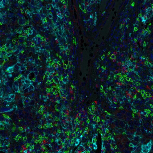
Detection of human CD8 (green) TIGIT (red) and PD-L1 (cyan) in breast carcinoma by IHC-IF. Antibodies: Rabbit anti-CD8 recombinant monoclonal [BLR044F] (A700-044), rabbit anti-TIGIT recombinant monoclonal [BLR047F] (A700-047) and rabbit anti-PD-L1 recombinant monoclonal [BLR020E] (A700-020). Secondary: HRP-conjugated goat anti-rabbit IgG (A120-501P). Substrate: Opal™ 520, Opal™ 690 and Opal™ 480. Counterstain: DAPI (blue).

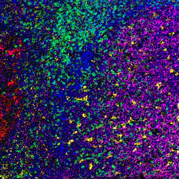
Detection of human CD3 (green), CD20 (magenta), CD68 (yellow), and cytokeratin (red) in FFPE tonsil by IHC-IF. Antibodies: Rabbit anti-CD3E recombinant monoclonal [BL-298-5D12] (A700-016), mouse anti-CD20 monoclonal [L26] (A500-017A), mouse anti-CD68 monoclonal [KP-1] (A500-018A), and mouse anti-Cytokeratin [AE1/AE3] (A500-019A). Secondary: HRP-conjugated goat anti-rabbit IgG (A120-501P) and HRP-conjugated goat anti-mouse IgG (A90-116P). Substrate: Opal™. Counterstain: DAPI (blue).

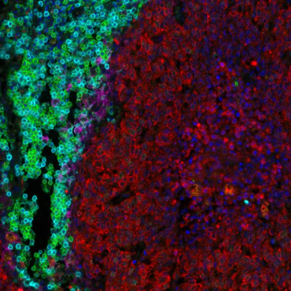
Detection of human CD3 (aqua), CD8 (green), cytokeratin (red), and PD-L1 (magenta) in FFPE lung carcinoma by IHC-IF. Rabbit anti-CD3E recombinant monoclonal [BL-298-5D12] (A700-016), rabbit anti-CD8 alpha [BLR044F] (A700-044), mouse anti-Cytokeratin [AE1/AE3] (A500-019A), and rabbit anti-PD-L1 [BLR020E] (A700-020). Secondary: HRP-conjugated goat anti-rabbit IgG (A120-501P) and HRP-conjugated goat anti-mouse IgG (A90-116P). Substrate: Opal™. Counterstain: DAPI (blue).