


Bethyl Laboratories
Rabbit anti-COP1/RFWD2 Antibody Affinity Purified
Primary
Catalog #A300-894A
TargetCOP1/RFWD2
HostRabbit
ClonalityPolyclonal
ReactivityMouse, Human
ApplicationIP, WB
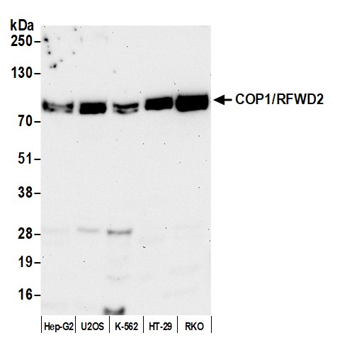
Image 1 of 3
local_shipping
Order within 5 hours and 31 minutes for shipment by today
Product Description
Constitutive photomorphogenic protein 1 (COP1) has been characterized in plants as a RING-finger-containing protein that functions to repress plant photomorphogenesis. In mammals, COP1 has been identified as an E3 ubiquitin ligase that negatively regulates p53 protein levels in unstressed cells. In response to DNA damage COP1 is phosphorylated by the ataxia telangiectasia mutated (ATM) protein kinase and is rapidly degraded releasing p53 to exert its tumor suppressor activities.
Specifications
Clonality
Polyclonal
Host
Rabbit
Immunogen
Between 1 and 50
Isotype
IgG
Reactivity
Mouse, Human
Format
Whole IgG
Conjugate Type
Unconjugated
Purity
Antigen Affinity Purified
Concentration
1000 µg/ml
Target Identity
TargetCOP1/RFWD2
Antigen Species
Human
Gene ID64326
Gene Symbol
COP1
Gene Name
ring finger and WD repeat domain 2
Uniprot IDQ8NHY2
Protein Name
E3 ubiquitin-protein ligase RFWD2
Alternate Names
constitutive photomorphogenesis protein 1 homolog • constitutive photomorphogenic protein 1 • E3 ubiquitin-protein ligase COP1 • E3 ubiquitin-protein ligase RFWD2 • hCOP1 • putative ubiquitin ligase COP1 • RFWD2 • ring finger and WD repeat domain 2, E3 ubiquitin protein ligase • RING finger and WD repeat domain protein 2 • RING finger protein 200 • RING-type E3 ubiquitin transferase RFWD2 • RNF200
Product Information
Applications
IP, WB
Buffer
Tris-buffered Saline containing 0.09% Sodium Azide, BSA-Free
Storage Conditions
2 - 8 °C
Physical State
Liquid
Shelf Life
1 year from date of receipt
Country of Origin
USA
Production & Epitope
Antibody was affinity purified using an epitope specific to COP1/RFWD2 immobilized on solid support.
The epitope recognized by A300-894A maps to a region between residue 1 and 50 of human Constitutive Photomorphogenic Protein-1/Ring Finger and WD Repeat Domain 2 using the numbering given in entry NP_071902.2 (GeneID 64326).
Immunoglobulin concentration was determined using Beer’s Law where 1mg/mL IgG has an A280 of 1.4.
Applications
All western blot analysis is performed using 5% Milk-TBST for blocking and as antibody diluent. Primary antibody is incubated overnight.
Western blots of cell lysates are performed using Goat anti-Rabbit IgG Heavy and Light Chain Antibody (A120-101P).
Western blots of immunoprecipitates are performed using an anti-Rabbit IgG Light Chain HRP-conjugated antibody with 5% Normal Pig Serum (Cat. No. S100-020) added to the blocking buffer.
Immunoprecipitation (IP)6 µg/mg lysate
Western Blot (WB)1:2000 - 1:10,000
Documents
Product DatasheetPDF · A300-894A · Lot 5 (current lot)Open
Product DatasheetPDF · A300-894A · Lot 5 (current lot)Open
Safety Data SheetPDF · A300-894AOpen
Citations / Publications
Services
Antibody Conjugation
Antibody and conjugate formats developed together, built to your specification.
Request CustomizationCustom Monoclonal Antibody Discovery Services
Full custom monoclonal development, from immunization through validation.
Explore Monoclonal DevelopmentVHH Discovery
Single-domain antibodies for structurally difficult targets a conventional monoclonal can't reach.
Explore VHH DiscoveryResources



1