


Bethyl Laboratories
Rabbit anti-VE-Cadherin Recombinant Monoclonal Antibody [BLR091G]
Monoclonal Antibodies
Catalog #A700-091
TargetVE-Cadherin
HostRabbit
CloneBLR091G
ReactivityHuman
ApplicationICC, IHC, IHC-IF, IP, WB
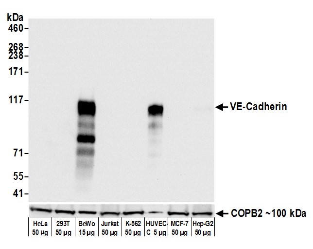
Image 1 of 5
local_shipping
Order within 10 hours and 33 minutes for shipment by Tuesday, November 10
Product Description
Cadherins are calcium-dependent cell adhesion proteins that preferentially interact with themselves in a homophilic manner in adjacent cells. VE-Cadherin, or Cadherin-5 plays an important role in endothelial cell biology through control of the cohesion and organization of the intercellular junctions. It associates with alpha-catenin forming a link to the cytoskeleton and acts in concert with KRIT1 and MPP5 to establish and maintain correct endothelial cell polarity and vascular lumen. [taken from the Universal Protein Resource (UniProt) www.uniprot.org/uniprot/P33151].
Specifications
Clonality
Recombinant Monoclonal
Clone
BLR091G
Host
Rabbit
Immunogen
Residues 48-599 (ECD)
Isotype
IgG
Reactivity
Human
Format
Whole IgG
Conjugate Type
Unconjugated
Purity
Purified
Concentration
1000 µg/ml
Target Identity
TargetVE-Cadherin
Antigen Species
Human
Gene ID1003
Gene Symbol
CDH5
Gene Name
cadherin 5
Uniprot IDP33151
Protein Name
Cadherin-5
Product Information
Applications
ICC, IHC, IHC-IF, IP, WB
Buffer
Borate Buffered Saline (BBS) pH 8.2 with 0.09% Sodium Azide, BSA-Free
Storage Conditions
2 - 8 °C
Physical State
Liquid
Shelf Life
1 year from date of receipt
Country of Origin
USA
Production & Epitope
Recombinant antibody was purified from cell culture supernatant.
Immunogen was a recombinant protein representing the extracellular domain of human VE-Cadherin (residues 48-599, NP_001786.2).
Applications
All western blot analysis is performed using 5% Milk-TBST for blocking and as antibody diluent. Primary antibody is incubated overnight.
Western blots of cell lysates are performed using Goat anti-Rabbit IgG Heavy and Light Chain Antibody (A120-101P).
Western blots of immunoprecipitates are performed using an anti-Rabbit IgG Light Chain HRP-conjugated antibody with 5% Normal Pig Serum (Cat. No. S100-020) added to the blocking buffer.
Immunocytochemistry (ICC)1:100 to 1:500. Epitope retrieval with citrate buffer pH6.0 is recommended for FFPE cell sections.
Immunohistochemistry (IHC)1:100 to 1:500. Epitope retrieval with citrate buffer pH6.0 is recommended for FFPE tissue sections.
Immunohistochemistry-Immunofluoresence (IHC-IF)1:100 to 1:500. Epitope retrieval with citrate buffer pH6.0 is recommended for FFPE tissue sections.
Immunoprecipitation (IP)6 µl/mg lysate
Western Blot (WB)1:1000
Documents
Product DatasheetPDF · A700-091 · Lot 1 (current lot)Open
Product DatasheetPDF · A700-091 · Lot 1 (current lot)Open
Safety Data SheetPDF · A700-091Open
Services
Antibody Conjugation
Antibody and conjugate formats developed together, built to your specification.
Request CustomizationCustom Monoclonal Antibody Discovery Services
Full custom monoclonal development, from immunization through validation.
Explore Monoclonal DevelopmentVHH Discovery
Single-domain antibodies for structurally difficult targets a conventional monoclonal can't reach.
Explore VHH DiscoveryResources



1