


Bethyl Laboratories
Rabbit anti-ARID1B Antibody, Affinity Purified
Primary
Catalog #A301-046A
TargetARID1B
HostRabbit
ClonalityPolyclonal
ReactivityHuman
ApplicationIHC, IP, WB
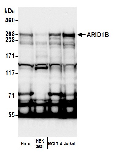
Image 1 of 3
local_shipping
Order within 2 hours and 12 minutes for shipment by today
Product Description
The SWI/SNF-related, matrix-associated, actin-dependent regulators of chromatin (SMARC), also called BRG1-associated factors (BAFs), have been identified as components of the human SWI/SNF-like chromatin-remodeling protein complexes. SWI/SNF complexes possess ATPase and DNA helicase activity that promotes transcriptional activation of genes normally repressed by chromatin structure. ARID1B is a noncatalytic subunit that is a member of the ARID (AT-rich interactive domain-containing protein) family of DNA binding proteins. ARID1B is encoded by a gene independent from ARID1A and exhibits functionally distinct characteristics.
Specifications
Clonality
Polyclonal
Host
Rabbit
Immunogen
Between 1400 and 1450
Isotype
IgG
Reactivity
Human
Format
Whole IgG
Conjugate Type
Unconjugated
Purity
Antigen Affinity Purified
Concentration
1000 µg/ml
Target Identity
TargetARID1B
Antigen Species
Human
Gene ID57492
Gene Symbol
ARID1B
Gene Name
AT-rich interaction domain 1B
Uniprot IDQ8NFD5
Protein Name
AT-rich interactive domain-containing protein 1B
Alternate Names
6A3-5 • ARID domain-containing protein 1B • AT rich interactive domain 1B (SWI1-like) • AT-rich interactive domain-containing protein 1B • BAF250B • BRG1-associated factor 250b • BRG1-binding protein ELD/OSA1 • BRG1-binding protein hELD/OSA1 • BRIGHT • CSS1 • DAN15 • ELD (eyelid)/OSA protein • ELD/OSA1 • hOsa2 • MRD12 • Osa homolog 2 • OSA2 • P250R
Product Information
Applications
IHC, IP, WB
Buffer
Tris-citrate/phosphate buffer, pH 7 to 8 containing 0.09% Sodium Azide
Storage Conditions
2 - 8 °C
Physical State
Liquid
Shelf Life
1 year from date of receipt
Country of Origin
USA
Production & Epitope
Antibody was affinity purified using an epitope specific to ARID1B immobilized on solid support.
The epitope recognized by A301-046A maps to a region between residue 1300 and 1350 of human AT rich interactive domain 1B using the numbering given in entry NP_059989.2 (GeneID 57492).
Immunoglobulin concentration was determined using Beer’s Law where 1mg/mL IgG has an A280 of 1.4.
Applications
All western blot analysis is performed using 5% Milk-TBST for blocking and as antibody diluent. Primary antibody is incubated overnight.
Western blots of cell lysates are performed using Goat anti-Rabbit IgG Heavy and Light Chain Antibody (A120-101P).
Western blots of immunoprecipitates are performed using an anti-Rabbit IgG Light Chain HRP-conjugated antibody with 5% Normal Pig Serum (Cat. No. S100-020) added to the blocking buffer.
Western Blot (WB)1:2000 - 1:10,000 (3-8% Tris-acetate gel recommended)
Immunoprecipitation (IP)6 µg/mg lysate
Immunohistochemistry (IHC)1:500 to 1:2000. Epitope retrieval with citrate buffer pH6.0 is recommended for FFPE tissue sections.
Documents
Product DatasheetPDF · A301-046A · Lot 4 (current lot)Open
Product DatasheetPDF · A301-046A · Lot 4 (current lot)Open
Safety Data SheetPDF · A301-046AOpen
Citations / Publications
Services
Antibody Conjugation
Antibody and conjugate formats developed together, built to your specification.
Request CustomizationCustom Monoclonal Antibody Discovery Services
Full custom monoclonal development, from immunization through validation.
Explore Monoclonal DevelopmentVHH Discovery
Single-domain antibodies for structurally difficult targets a conventional monoclonal can't reach.
Explore VHH DiscoveryResources



1