


Bethyl Laboratories
Rabbit anti-CSN3 Antibody Affinity Purified
Primary
Catalog #A300-029A
TargetCSN3
HostRabbit
ClonalityPolyclonal
ReactivityHuman
ApplicationWB
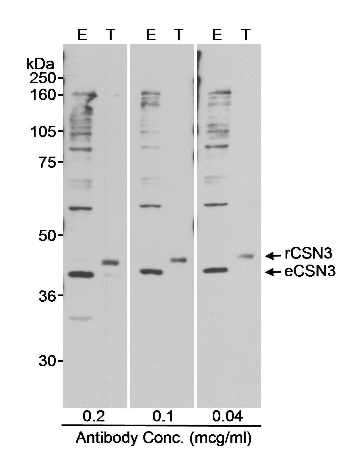
Image 1 of 1
local_shipping
Order within 12 hours and 53 minutes for shipment by Tuesday, November 10
Product Description
CSN3 is one of eight subunits (CSN1 to 8) of the highly conserved COP9 signalsome (CSN) complex originally identified as a regulator of light-mediated development in Arabidopsis. Characterization of CSN from yeast to mammals reveals its function as a modulator of signal transduction pathways involved in a variety of cellular and developmental processes. One of the major functions of the CSN is the regulation of protein degradation via intersection with the ubiquitin-proteasome pathway and regulation of E3-ubiquitin ligases. The CSN also possesses kinase activity. CSN3 is the kinase associated with the CSN complex that phosphorylates signal transducers such as I-kappa-Balpha, p105, and c-Jun. Human CSN3 maps to the Smith-Magenis syndrome locus. Microdeletions in this locus results in mental retardation, skeletal abnormalities, sleep disorder, and neurobehavior anomalies.
Specifications
Clonality
Polyclonal
Host
Rabbit
Immunogen
Between 350 and 400
Isotype
IgG
Reactivity
Human
Format
Whole IgG
Conjugate Type
Unconjugated
Purity
Antigen Affinity Purified
Concentration
1000 µg/ml
Target Identity
TargetCSN3
Antigen Species
Human
Gene ID8533
Gene Symbol
COPS3
Gene Name
COP9 signalosome subunit 3
Uniprot IDQ9UNS2
Protein Name
COP9 signalosome complex subunit 3
Product Information
Applications
WB
Buffer
Tris-citrate/phosphate buffer, pH 7 to 8 containing 0.09% Sodium Azide
Storage Conditions
2 - 8 °C
Physical State
Liquid
Shelf Life
1 year from date of receipt
Country of Origin
USA
Production & Epitope
Antibody was affinity purified using an epitope specific to CSN3 immobilized on solid support.
The epitope recognized by A300-029A maps to a region between residues 325 and 375 of human COP9 constitutive photomorphogenic homolog subunit 3 using the numbering given in entry NP_003644.2 (GeneID 8533).
Immunoglobulin concentration was determined using Beer’s Law where 1mg/mL IgG has an A280 of 1.4.
Applications
All western blot analysis is performed using 5% Milk-TBST for blocking and as antibody diluent. Primary antibody is incubated overnight.
Western blots of cell lysates are performed using Goat anti-Rabbit IgG Heavy and Light Chain Antibody (A120-101P).
Western Blot (WB)1:2000 - 1:20,000
Documents
Product DatasheetPDF · A300-029A · Lot 1 (current lot)Open
Product DatasheetPDF · A300-029A · Lot 1 (current lot)Open
Safety Data SheetPDF · A300-029AOpen
Services
Antibody Conjugation
Antibody and conjugate formats developed together, built to your specification.
Request CustomizationCustom Monoclonal Antibody Discovery Services
Full custom monoclonal development, from immunization through validation.
Explore Monoclonal DevelopmentVHH Discovery
Single-domain antibodies for structurally difficult targets a conventional monoclonal can't reach.
Explore VHH DiscoveryResources



1