


Bethyl Laboratories
Rabbit anti-NUMA IHC Antibody Affinity Purified
Primary
Catalog #IHC-00302
TargetNUMA
HostRabbit
ClonalityPolyclonal
ReactivityHuman
ApplicationIHC, IHC-IF
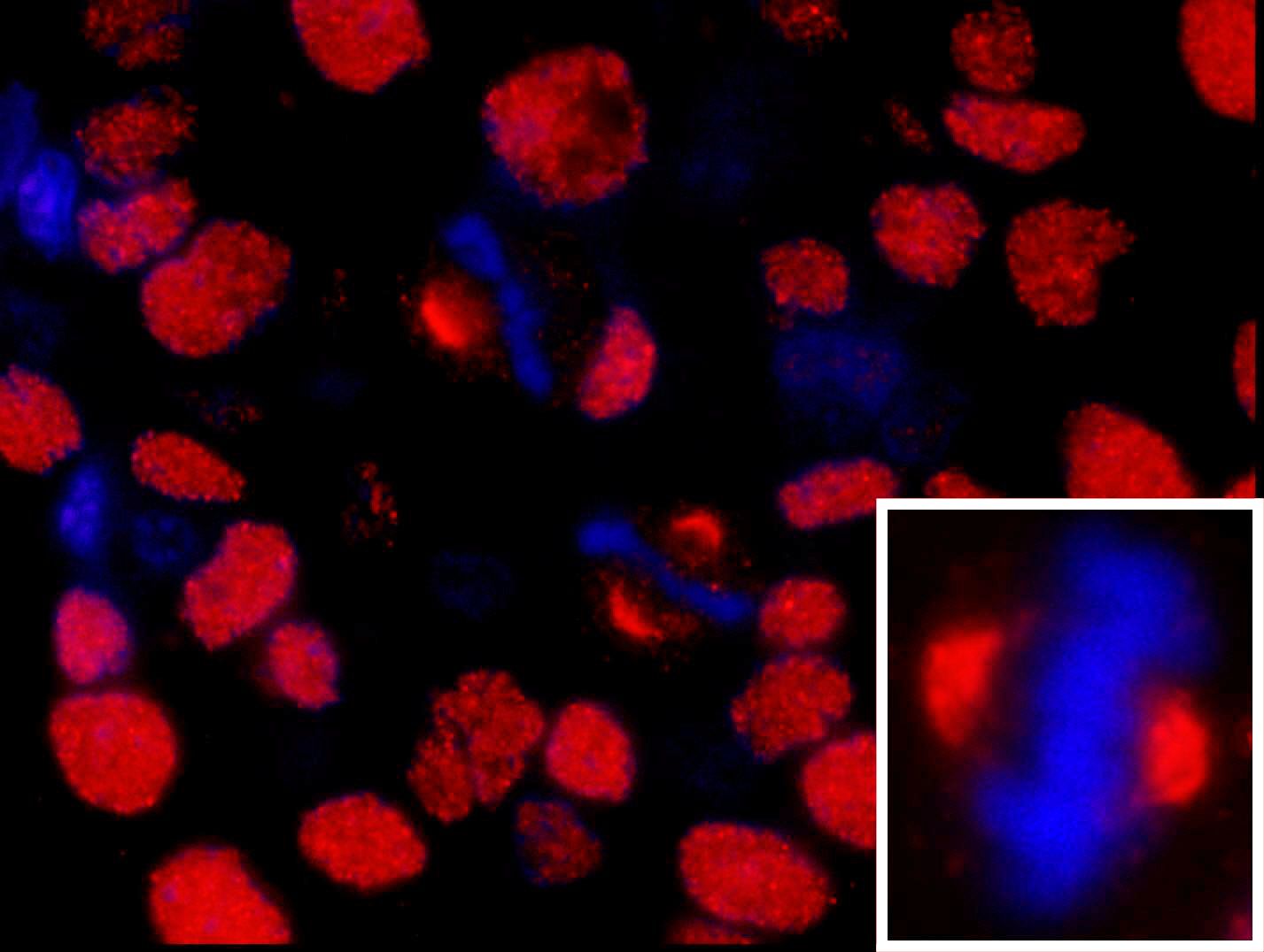
Image 1 of 1
local_shipping
Order within 6 hours and 31 minutes for shipment by today
Product Description
The nuclear mitotic apparatus protein (NuMA) is a critical component of the mitotic spindle that is responsible for organizing minus-ends of microtubules at spindle poles. NUMA is regulated by phosphorylation and associates with dynein and dynactin in its phosphorylated form. Dephosphorylation of NUMA in anaphase results in the release from dynein and dynactin to allow spindle disassembly. NUMA, in association with dynein and dynactin, also interacts with 4.1R in a complex of spindle pole organizing proteins. Other NUMA interacting proteins include GAS41, tankyrase, and Rae1. These interactions indicate a broader role for NUMA function in the nucleus.
Specifications
Clonality
Polyclonal
Host
Rabbit
Immunogen
Between 925 and 975
Isotype
IgG
Reactivity
Human
Format
Whole IgG
Conjugate Type
Unconjugated
Purity
Antigen Affinity Purified
Concentration
50 µg/ml
Target Identity
TargetNUMA
Antigen Species
Human
Gene ID4926
Gene Symbol
NUMA1
Gene Name
nuclear mitotic apparatus protein 1
Uniprot IDQ14980
Protein Name
Nuclear mitotic apparatus protein 1
Product Information
Applications
IHC, IHC-IF
Buffer
Tris-buffered Saline containing 0.1% BSA and 0.09% Sodium Azide
Storage Conditions
2 - 8 °C
Physical State
Liquid
Shelf Life
1 year from date of receipt
Country of Origin
USA
Production & Epitope
Antibody was affinity purified using an epitope specific to NUMA immobilized on solid support.
The epitope recognized by IHC-00302 maps to a region between residue 900 and 950 of human nuclear mitotic apparatus protein 1 using the numbering given in entry NP_006176.2 (GeneID 4926).
Immunoglobulin concentration was determined using Beer’s Law where 1mg/mL IgG has an A280 of 1.4.
Applications
Epitope exposure is recommended.
Epitope exposure with citrate buffer will enhance staining.
Likely to work with frozen sections.
In some cases, the antibody may be diluted further than indicated.
Immunohistochemistry (IHC)1:100 - 1:500
Immunohistochemistry-Immunofluoresence (IHC-IF)1:50 - 1:500
Documents
Safety Data SheetPDF · IHC-00302Open
Product DatasheetPDF · IHC-00302 · Lot 1 (current lot)Open
Product DatasheetPDF · IHC-00302 · Lot 1 (current lot)Open
Services
Antibody Conjugation
Antibody and conjugate formats developed together, built to your specification.
Request CustomizationCustom Monoclonal Antibody Discovery Services
Full custom monoclonal development, from immunization through validation.
Explore Monoclonal DevelopmentVHH Discovery
Single-domain antibodies for structurally difficult targets a conventional monoclonal can't reach.
Explore VHH DiscoveryResources



1