


Bethyl Laboratories
Rabbit anti-PSMA1 Antibody Affinity Purified
Primary
Catalog #A303-846A
TargetPSMA1
HostRabbit
ClonalityPolyclonal
ReactivityHuman
ApplicationIHC, IP, WB
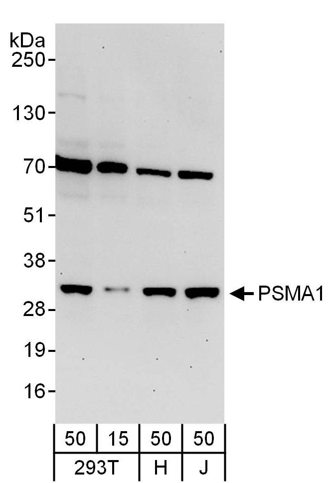
Image 1 of 3
local_shipping
Order within 6 hours and 21 minutes for shipment by Tuesday, November 24
Product Description
PSMA1 [proteasome (prosome, macropain) subunit, alpha type, 1] is a subunit of the proteasome, a multicatalytic proteinase complex with a highly ordered ring-shaped 20S core structure. The core structure is composed of 4 rings of 28 non-identical subunits; 2 rings are composed of 7 alpha subunits and 2 rings are composed of 7 beta subunits. Proteasomes are distributed throughout eukaryotic cells at a high concentration and cleave peptides in an ATP/ubiquitin-dependent process in a non-lysosomal pathway. An essential function of a modified proteasome, the immunoproteasome, is the processing of class I MHC peptides. PSMA1 is a member of the peptidase T1A family that is a 20S core alpha subunit [taken from NCBI Entrez Gene (Gene ID: 5682)].
Specifications
Clonality
Polyclonal
Host
Rabbit
Immunogen
Between 213 and C-terminus
Isotype
IgG
Reactivity
Human
Format
Whole IgG
Conjugate Type
Unconjugated
Purity
Antigen Affinity Purified
Concentration
1000 µg/ml
Target Identity
TargetPSMA1
Antigen Species
Human
Gene ID5682
Gene Symbol
PSMA1
Gene Name
proteasome subunit alpha 1
Uniprot IDP25786
Protein Name
Proteasome subunit alpha type-1
Alternate Names
30 kDa prosomal protein • epididymis secretory protein Li 275 • HC2 • HEL-S-275 • macropain subunit C2 • macropain subunit nu • multicatalytic endopeptidase complex subunit C2 • NU • PROS30 • PROS-30 • proteasome (prosome, macropain) subunit, alpha type, 1 • proteasome component C2 • proteasome nu chain • proteasome subunit alpha type-1 • proteasome subunit nu • proteasome subunit, alpha-type, 1 • protein P30-33K • testicular tissue protein Li 150
Product Information
Applications
IHC, IP, WB
Buffer
Tris-citrate/phosphate buffer, pH 7 to 8 containing 0.09% Sodium Azide
Storage Conditions
2 - 8 °C
Physical State
Liquid
Shelf Life
1 year from date of receipt
Country of Origin
USA
Production & Epitope
Antibody was affinity purified using an epitope specific to PSMA1 immobilized on solid support.
The epitope recognized by A303-846A maps to a region between residue 213 and 263 of human Proteasome (Prosome, Macropain) Subunit, Alpha Type, 1 using the numbering given in entry NP_002777.1 (GeneID 5682).
Immunoglobulin concentration was determined using Beer’s Law where 1mg/mL IgG has an A280 of 1.4.
Applications
All western blot analysis is performed using 5% Milk-TBST for blocking and as antibody diluent. Primary antibody is incubated overnight.
Western blots of cell lysates are performed using Goat anti-Rabbit IgG Heavy and Light Chain Antibody (A120-101P).
Western blots of immunoprecipitates are performed using an anti-Rabbit IgG Light Chain HRP-conjugated antibody with 5% Normal Pig Serum (Cat. No. S100-020) added to the blocking buffer.
Immunohistochemistry (IHC)1:500 - 1:2000. Epitope retrieval with citrate buffer pH 6.0 is recommended for FFPE tissue sections.
Immunoprecipitation (IP)6 µg/mg lysate
Western Blot (WB)1:2000 - 1:10,000
Documents
Product DatasheetPDF · A303-846A · Lot 1 (current lot)Open
Product DatasheetPDF · A303-846A · Lot 1 (current lot)Open
Safety Data SheetPDF · A303-846AOpen
Services
Antibody Conjugation
Antibody and conjugate formats developed together, built to your specification.
Request CustomizationCustom Monoclonal Antibody Discovery Services
Full custom monoclonal development, from immunization through validation.
Explore Monoclonal DevelopmentVHH Discovery
Single-domain antibodies for structurally difficult targets a conventional monoclonal can't reach.
Explore VHH DiscoveryResources



1