
Bethyl Laboratories
Rabbit anti-Ki-67 Recombinant Monoclonal Antibody [BLR021E]
Monoclonal Antibodies
Catalog #A700-021
TargetKi-67
HostRabbit
CloneBLR021E
ReactivityHuman
ApplicationFlow Cyt, ICC, ICC-IF, IHC, IHC-IF, mIF
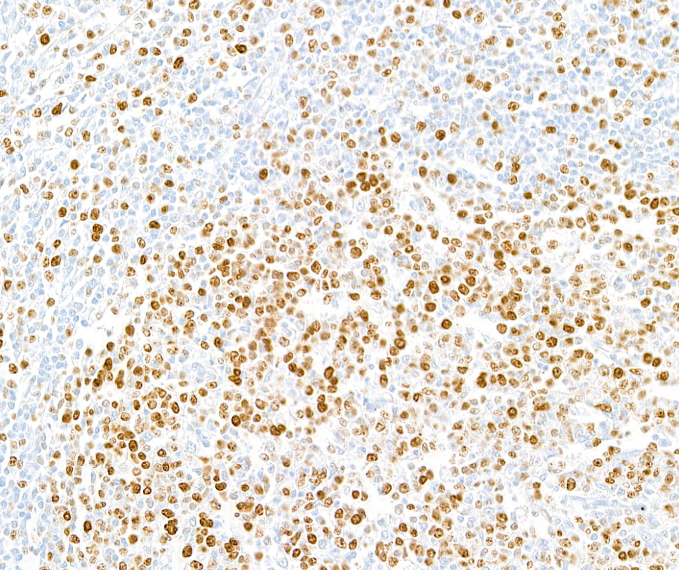
Image 1 of 4
local_shipping
Order within 23 hours and 11 minutes for shipment by today
Product Description
Ki-67 is a marker of cell proliferation detected in cells that are in G1-, S-, G2-, and M-phase of the cell cycle and absent in cells in G0. Ki-67 is detected exclusively in the nucleus, and its detection by immunohistological methods has proven to have diagnostic and prognostic value in the study of a variety of human tumors. The specific cellular functions of Ki-67 have not been elucidated, but it is proposed to function in the organization and maintenance of the architecture of DNA and in the synthesis of ribosomes during mitosis.
Specifications
Clonality
Recombinant Monoclonal
Clone
BLR021E
Host
Rabbit
Immunogen
Between 1200 and 1250
Isotype
IgG
Reactivity
Human
Format
Whole IgG
Conjugate Type
Unconjugated
Purity
Purified
Concentration
50 µg/ml
Target Identity
TargetKi-67
Antigen Species
Human
Gene ID4288
Gene Symbol
MKI67
Gene Name
marker of proliferation Ki-67
Uniprot IDP46013
Protein Name
Antigen KI-67
Product Information
Applications
Flow Cyt, ICC, ICC-IF, IHC, IHC-IF, mIF
Buffer
Borate Buffered Saline (BBS) pH 8.2 with 0.1% BSA and 0.09% Sodium Azide
Storage Conditions
2 - 8 °C
Physical State
Liquid
Shelf Life
1 year from date of receipt
Country of Origin
USA
Production & Epitope
Recombinant antibody was purified from cell culture supernatant.
Immunogen was a peptide representing the repetitive 66 bp element of human Ki-67 using the numbering given in entry NP_002408.2 (GeneID 4288).
Applications
Flow Cytometry (Flow Cyt)Fixed in 4% formaldehyde and permeabilized with 90% methanol. 1 µl per 1 x 10^6 cells.
Immunocytochemistry (ICC)1:100 to 1:500. Epitope retrieval with citrate buffer pH 6.0 is recommended for FFPE cell sections.
Immunocytochemistry-Immunofluoresence (ICC-IF)1:100 - 1:500
Immunohistochemistry (IHC)1:100 to 1:500. Epitope retrieval with citrate buffer pH 6.0 is recommended for FFPE tissue sections.
Immunohistochemistry-Immunofluoresence (IHC-IF)1:100 to 1:500. Epitope retrieval with citrate buffer pH6.0 is recommended for FFPE tissue sections.
Multiplex Immunofluorescence (mIF)1:250
Documents
Product DatasheetPDF · A700-021 · Lot 3 (current lot)Open
Product DatasheetPDF · A700-021 · Lot 3 (current lot)Open
Product DatasheetPDF · A700-021 · Lot 231213Open
Safety Data SheetPDF · A700-021Open
Related Products
Services
Antibody Conjugation
Antibody and conjugate formats developed together, built to your specification.
Request Customization
Custom Monoclonal Antibody Discovery Services
Full custom monoclonal development, from immunization through validation.
Explore Monoclonal Development
VHH Discovery
Single-domain antibodies for structurally difficult targets a conventional monoclonal can't reach.
Explore VHH Discovery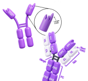
Resources

Standard Western Blot Protocol
Read the Protocol
Immunoprecipitation Protocol
Read the Protocol
Simple Western™ Resources
Explore Simple Western™
1