
Bethyl Laboratories
Rabbit anti-TIM3 Recombinant Monoclonal Antibody [BLR033F]
Monoclonal Antibodies
Catalog #A700-033
TargetTIM3
HostRabbit
CloneBLR033F
ReactivityHuman
ApplicationFlow Cyt, ICC, IHC, IHC-IF, WB

Image 1 of 5
local_shipping
Order within 20 hours and 33 minutes for shipment by Tuesday, November 17
Product Description
TIM3 (HAVCR2) is a cell surface receptor implicated in modulating innate and adaptive immune responses. It is generally accepted to have an inhibiting function. Reports on stimulating functions suggest that the activity may be influenced by the cellular context and/or the respective ligand. TIM3 regulates macrophage activation, inhibits T-helper type 1 lymphocyte (Th1)-mediated auto- and alloimmune responses, and promotes immunological tolerance. In CD8+ cells TIM3 attenuates TCR-induced signaling, specifically by blocking NF-kappaB and NFAT promoter activities resulting in the loss of IL-2 secretion. [taken from the Universal Protein Resource (UniProt) www.uniprot.org/uniprot/Q8TDQ0].
Specifications
Clonality
Recombinant Monoclonal
Clone
BLR033F
Host
Rabbit
Immunogen
Residues 22-201 (ECD)
Isotype
IgG
Reactivity
Human
Format
Whole IgG
Conjugate Type
Unconjugated
Purity
Purified
Concentration
1000 µg/ml
Target Identity
TargetTIM3
Antigen Species
Human
Gene ID84868
Gene Symbol
HAVCR2
Gene Name
hepatitis A virus cellular receptor 2
Uniprot IDQ8TDQ0
Protein Name
Hepatitis A virus cellular receptor 2
Alternate Names
CD antigen CD366 • CD366 • HAVcr-2 • hepatitis A virus cellular receptor 2 • kidney injury molecule-3 • KIM-3 • T cell immunoglobulin mucin 3 • T-cell immunoglobulin and mucin domain-containing protein 3 • T-cell immunoglobulin mucin family member 3 • T-cell immunoglobulin mucin receptor 3 • T-cell membrane protein 3 • TIM3 • Tim-3 • TIMD3 • TIMD-3
Product Information
Applications
Flow Cyt, ICC, IHC, IHC-IF, WB
Buffer
Borate Buffered Saline (BBS) pH 8.2 with 0.09% Sodium Azide, BSA-Free
Storage Conditions
2 - 8 °C
Physical State
Liquid
Shelf Life
1 year from date of receipt
Country of Origin
USA
Production & Epitope
Recombinant antibody was purified from cell culture supernatant.
Immunogen was a recombinant protein representing the extracellular domain of human TIM3 (HAVCR2) (residues 22-201, NP_116171.3).
Applications
All western blot analysis is performed using 5% Milk-TBST for blocking and as antibody diluent. Primary antibody is incubated overnight.
Western blots of cell lysates are performed using Goat anti-Rabbit IgG Heavy and Light Chain Antibody (A120-101P).
Flow Cytometry (Flow Cyt)Fixed in 4% formaldehyde and permeabilized with 90% methanol. 0.5 µl per 1 x 10^6 cells.
Immunocytochemistry (ICC)1:100 to 1:500. Epitope retrieval with citrate buffer pH6.0 is recommended for FFPE cell sections.
Immunohistochemistry (IHC)1:100 to 1:500. Epitope retrieval with citrate buffer pH6.0 is recommended for FFPE tissue sections.
Immunohistochemistry-Immunofluoresence (IHC-IF)1:100 to 1:500. Epitope retrieval with citrate buffer pH6.0 is recommended for FFPE tissue sections.
Western Blot (WB)1:1000
Documents
Product DatasheetPDF · A700-033 · Lot 2 (current lot)Open
Product DatasheetPDF · A700-033 · Lot 2 (current lot)Open
Safety Data SheetPDF · A700-033Open
Related Products
Services
Antibody Conjugation
Antibody and conjugate formats developed together, built to your specification.
Request Customization
Custom Monoclonal Antibody Discovery Services
Full custom monoclonal development, from immunization through validation.
Explore Monoclonal Development
VHH Discovery
Single-domain antibodies for structurally difficult targets a conventional monoclonal can't reach.
Explore VHH Discovery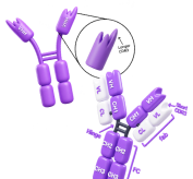
Resources

Standard Western Blot Protocol
Read the Protocol
Immunoprecipitation Protocol
Read the Protocol
Simple Western™ Resources
Explore Simple Western™
1